The School of Health Sciences from the Universidad Francisco de Vitoria (UFV) has incorporated 3D scanning and printing technologies into the teaching of Human Anatomy to reinforce their students’ hands-on learning. Through an activity conducted as part of the Occupational Therapy “ Bachelor's Degree ,” students worked with real skeletal structures to transform them into digital models and prepare them for reproduction via 3D printing—an experience that connects anatomical knowledge with the potential applications of these technologies in the healthcare field.
The initiative is part of the Human Anatomy I course and is based on an educational approach that seeks to integrate the scientific basis through experiential learning and the development of technological skills. In the Bachelor's Degree in Occupational Therapy at UFV, this experience serves as an introduction to technologies that students will later explore in courses such as “Prosthetics, Orthotics, and Assistive Devices in 3D Printing,” which is offered in the third year. In this way, the curriculum establishes a connection between the study of basic sciences and their future professional application.
From the study of anatomy to the creation of three-dimensional models
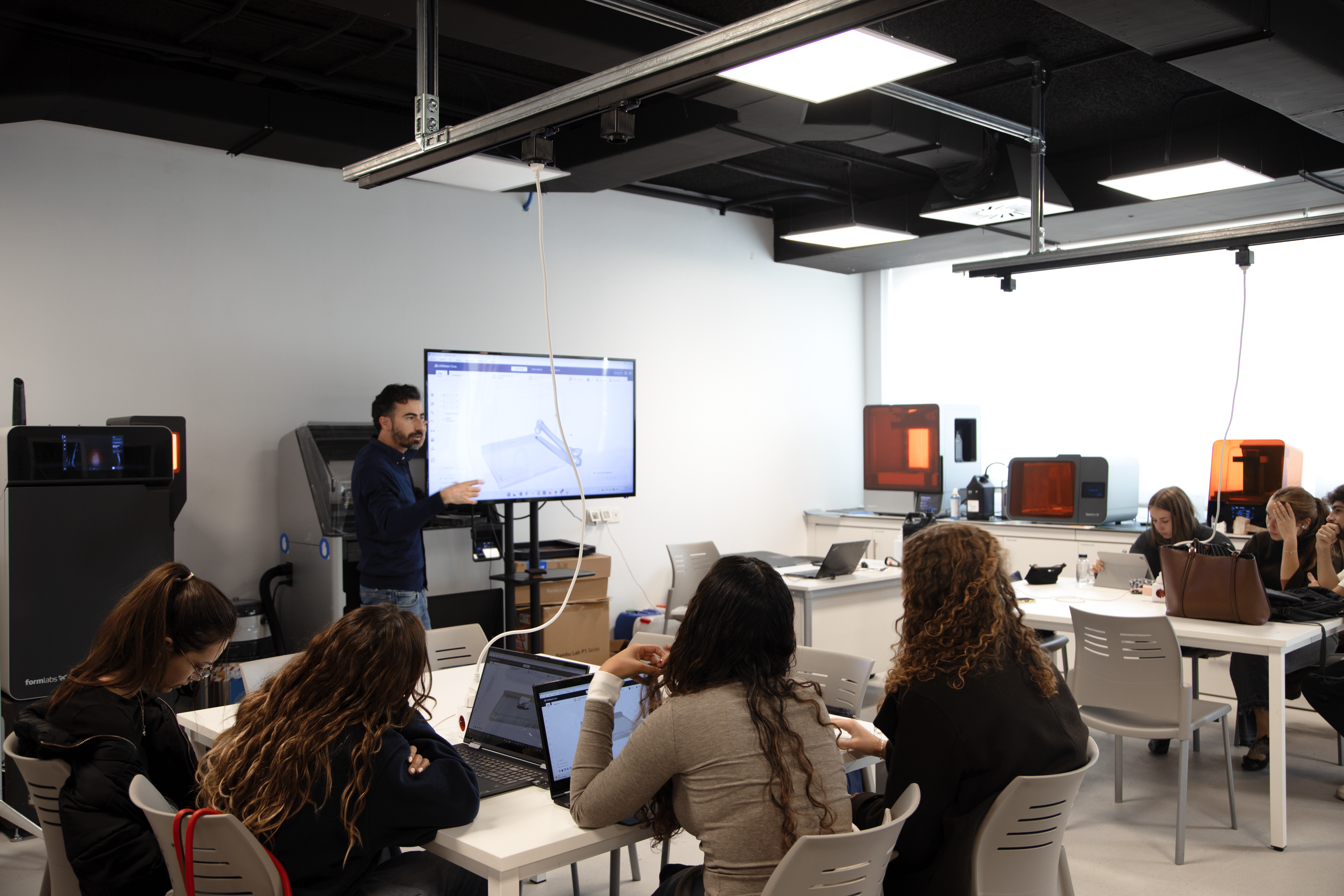
The activity was organized into two hands-on sessions. In the UFV Dissection Center, the students learned how to use 3D scanning tools to digitize various bone structures. This process allowed them to explore the anatomical structures from different perspectives and understand their spatial organization.
They also discussed the process of printing the previously scanned structures, thus completing a series of steps that allows students to transform an anatomical specimen into a reproducible physical model.
The professor teaching the course, África Ruiz, explains that this methodology encourages more active student participation: “In this activity, students don’t just observe an anatomical structure; they have to explore it, orient it correctly, identify its anatomical landmarks, scan it, and convert it into a digital model "which it then prepares for printing."
In addition, this experience is part of the course’s academic assessment. Students must prepare a group project on the structure they have scanned and printed, applying the theoretical knowledge acquired during the course and developing their ability to interpret the anatomical characteristics of each specimen.
Technology Applied to the Training of Future Occupational Therapists
One of the distinguishing features of this activity is its connection to the skills students will need in his future career. 3D printing offers possibilities for manufacturing and customizing assistive devices to suit each person's characteristics and needs, an area of particular interest to occupational therapy.
In this regard, África Ruiz points out that “for future occupational therapists It's particularly interesting understand the possibilities these technologies offer, since the manufacture and customization of objects through 3D printing can open up new possibilities for the development and adaptation of assistive products and other solutions aimed at specific needs "of people."
This experience also allows students to make connections between Human Anatomy I and other courses at the Bachelor's Degree related to movement, bodily function, and adaptation to the environment. In this way, students begin to understand how the scientific knowledge acquired during the first courses can be applied later to address actual needs.
A 3D anatomical repository to expand teaching resources
The project also calls for the creation of a digital repository of three-dimensional anatomical models based on the scanned structures by the students themselves. These files may be used in future classes and training activities, as well as physically reproduced to facilitate the study of anatomy.
This resource bank will make it possible to build on the work done in subsequent editions of the course and opens up the possibility of sharing the models with other degree programs in the School of Health Sciences that include the study of anatomy in their curricula.
The initiative also includes a process for evaluating the teaching experience to analyze students' perceptions of its usefulness and its contribution to learning. The results will make it possible to identify possible improvements and guide the development of future activities.
With this project, the UFV School of Health Sciences continues to advance a teaching model that integrates scientific rigour with the practical application of knowledge, preparing future healthcare professionals to understand the possibilities of innovation and put them to work for the benefit of people.